

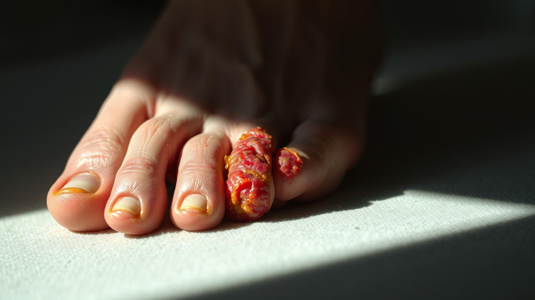
People with diabetes face a higher risk of foot problems. A foot ulcer affects many, and timely action cuts the chance of severe infections and hospital stays.
The earliest clues are often local: redness, warmth, swelling, tenderness, or pus. Clinicians diagnose a diabetes-related foot infection when two or more local findings are present.
Quick assessment by a team — including podiatry, vascular, and infectious disease specialists — improves outcomes and lowers amputation rates.
This guide will show readers how to spot warning changes at the foot, understand severity, and seek the right care. It also previews tests clinicians may use, like imaging, cultures after debridement, and lab markers.
Dominion Mobile Wound Care is featured throughout as a provider that brings expert evaluation and treatment directly to patients who need prompt, multidisciplinary support.
Key Takeaways
- Watch the foot closely for redness, warmth, swelling, tenderness, or purulent drainage.
- Two or more local findings usually indicate a clinical diagnosis that needs prompt care.
- Early, team-based evaluation reduces hospitalization and lowers amputation risk.
- Clinicians may use imaging, cultures, and labs to confirm spread and depth.
- Dominion Mobile Wound Care offers on-site assessment and coordinated treatment.
Why Early Detection Matters for Diabetic Foot Health in the United States
Early recognition of subtle foot changes can stop a minor problem from becoming a limb-threatening emergency.
Diabetes increases risk because loss of sensation, poor blood flow, and immune changes let small skin breaks turn into ulcers and then deeper infection.
About 40% of foot ulcers become infected, and more than 130,000 diabetes-related amputations occurred in the U.S. in 2016. Outcomes after amputation are serious, with high mortality at five years.
How disease factors raise risk
Major factors include peripheral neuropathy, peripheral arterial disease, and impaired immunity. Each factor makes healing slower and inflammation more likely.
The role of multidisciplinary care
Coordinated, on-site care from a team reduces hospitalization and lowers amputation rate by speeding diagnosis, grading, and treatment. The International Working Group offers validated grading to guide decisions.
- Regular foot care and annual exams help catch changes early.
- Early blood tests (white cells, ESR) and imaging speed accurate management.
- Dominion Mobile Wound Care provides on-site assessment to shorten time to treatment.
Who Is at Higher Risk: Peripheral Neuropathy, Peripheral Arterial Disease, and Immunity
When nerves fail and blood flow falls, small foot injuries can escalate quickly. Peripheral neuropathy causes loss of protective sensation for pain and temperature. About half of affected people feel no symptoms and can unknowingly injure the foot or toe.
Loss of protective sensation and unnoticed wounds
Neuropathy lets pressure points and minor cuts progress to ulcers. Daily checks of the skin, nails, and between toes help catch changes early. Dominion Mobile Wound Care prioritizes these high-risk patients for home monitoring and timely intervention.
Blood flow issues that slow healing and raise infection risk
Peripheral arterial disease limits blood delivery and delays healing. PAD affects roughly 10%–40% of people with diabetes. An ankle-brachial index under 0.9 suggests occlusive disease; under 0.5 indicates significant PAD that increases risk of poor outcomes and amputation.
- Highest risk: neuropathy, PAD, prior ulcer or amputation, and foot deformities.
- Vascular testing and ABI values guide revascularization decisions.
- Prompt reporting of any toe or foot skin change prevents escalation.
Recognizing diabetic wound infection signs at the wound site
Local changes at the foot can be the first clear cue that a problem needs prompt attention.
Local inflammation and temperature
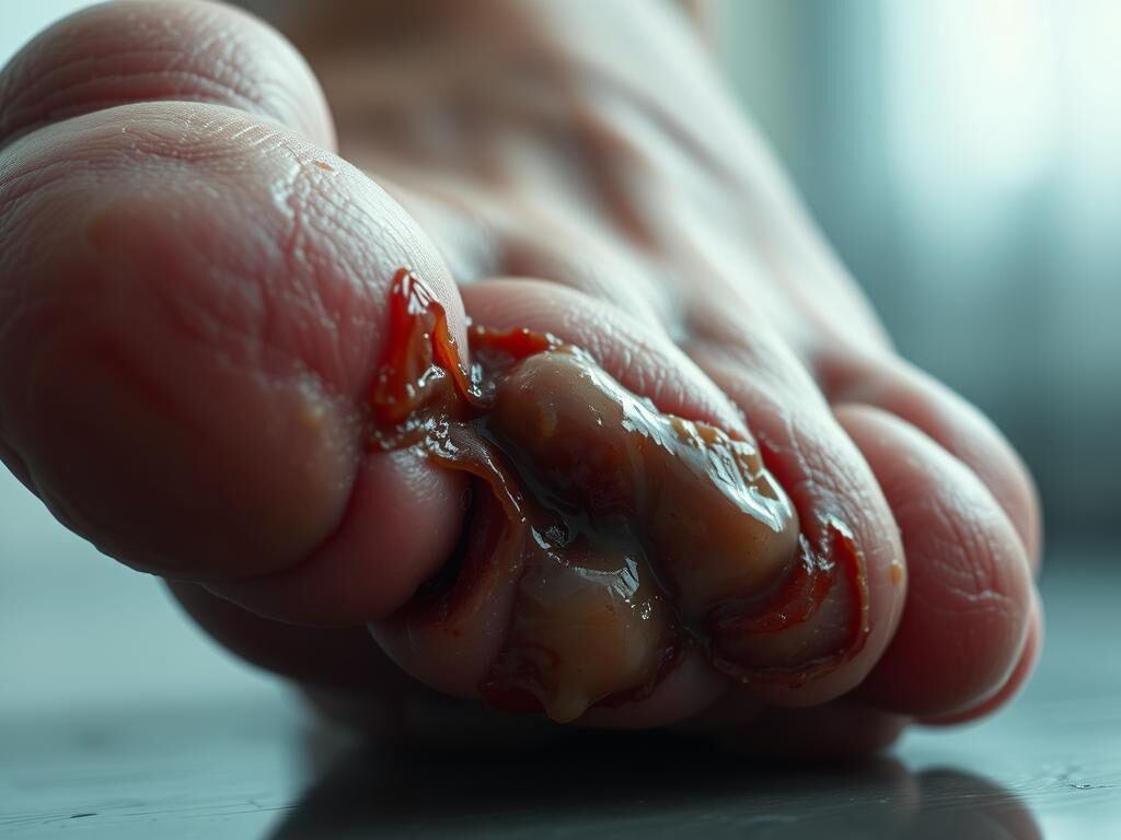
New or increasing redness, warmth, swelling, or tenderness around an ulcer or skin break usually signals local inflammation. A higher skin temperature compared with the other foot is an important objective clue.
Drainage and odor
Purulent drainage, foul odor, or a nonhealing area strongly point to active infection and merit professional review. Persistent drainage or a change in volume or color should be recorded and shown to a clinician.
Skin and tissue changes
Induration, necrotic or friable tissues, and spreading cellulitis around ulcers suggest deeper tissue involvement. Neuropathy may blunt pain, so lack of pain does not rule out a serious problem.
- Diagnosis usually requires at least two classic findings such as purulence or inflammation.
- Avoid sole reliance on superficial swabs; debrided base or deep tissue yields better cultures.
- Inspect between toes, under calluses, and at pressure points; document size, color, drainage, and odor.
Dominion Mobile Wound Care educates patients to spot these local changes early and can provide bedside assessment, debridement, and culture collection to guide treatment and help prevent spread to bone or joint.
How Severe Is It? Using IWGDF/IDSA Grading to Classify Infection
Clinicians use a standardized grading to turn bedside observations into a clear treatment plan. The international working group and IDSA offer a four-level scale that predicts hospitalization and amputation risk.
Uninfected and mild
Level 1 means no local problem at the site. Level 2, or mild, involves only skin or subcutaneous tissue. Erythema is small—greater than 0.5 cm but up to 2 cm. There are no systemic features.
Moderate
Moderate cases show erythema beyond 2 cm or involvement of deeper tissue such as tendon, muscle, joint, or bone. Abscess, septic arthritis, or osteomyelitis fit here. Imaging and prompt culture-guided management are usually needed.
Severe
Severe infection meets systemic inflammatory response thresholds: abnormal temperature, heart or respiratory rate, or white blood cell count derangement. These cases carry higher amputation rates and require urgent, often hospital-level, multidisciplinary treatment.
- Why this matters: Grading guides antibiotics, urgency of debridement, and need for vascular or surgical referral.
- Dominion Mobile Wound Care applies this scale at bedside to fast-track imaging, labs, and specialist transfer when needed.
Diagnosis Done Right: From Bedside Clues to Imaging and Labs
A stepwise approach—exam first, targeted testing next—yields the best diagnostic clarity.
Probe-to-bone testing and careful debridement for accurate cultures
After gentle sharp debridement, a probe-to-bone test can help detect bone involvement. When done after cleaning the area, its sensitivity is roughly 87% and specificity about 91% for osteomyelitis in outpatients.
Avoid superficial swabs. Deep tissue or bone cultures taken at the time of debridement better identify true pathogens and guide antibiotic choices.
When to order imaging: radiographs first, then magnetic resonance imaging if needed
Start with plain radiographs to look for gas, foreign material, or clear bony change. Early osteomyelitis may be missed on X-ray.
Magnetic resonance imaging is the most accurate next step—about 90% sensitive and 80% specific—and clarifies soft tissue extent and bone disease. Use CT if MRI is contraindicated.
Laboratory markers: white blood cell count, C-reactive protein, erythrocyte sedimentation rate
White blood cell counts can be normal; clinicians should not rely on them alone. An erythrocyte sedimentation rate above 70 mm/h supports bone infection, while C-reactive protein is useful to track response to treatment.
Dominion Mobile Wound Care coordinates on-site debridement, proper culture collection, and mobile imaging referrals. This reduces delays, improves antibiotic stewardship, and speeds recovery.
- Document ulcer depth, probe to bone at toes or prominences, and reassess after initial care.
- Consider comorbid disease and renal function when selecting imaging and lab pathways.
- Accurate diagnosis reduces unnecessary broad-spectrum therapy and shortens recovery time.
When to Seek Help Now: Red Flags That Warrant Urgent Care
When the foot shows sudden, aggressive changes, delays in care can lead to loss of limb or systemic illness. Quick recognition of urgent features shortens time to treatment like antibiotics, debridement, or revascularization.

Immediate red flags to act on
- Rapidly spreading redness, severe pain out of proportion, fever, foul odor, or visible bone at a toe or other bony site.
- New black tissue, gangrene, or crepitus suggesting necrosis or gas in tissue — these are emergencies.
- Nonhealing ulcers, sudden increase in drainage, or a marked rise in swelling that fails to improve with basic care.
- Cold foot, weak or absent pulses, and slow capillary refill—signs consistent with peripheral arterial disease (ABI <0.5 may indicate severe ischemia).
What to expect and when to call
Dominion Mobile Wound Care triages urgent symptoms rapidly and can direct patients to emergency services or arrange expedited specialty care. Any exposed bone, systemic illness, or crepitus should prompt immediate transfer to hospital-level care.
Failure to improve after initial treatment or worsening pain — even in people with loss of sensation — requires prompt re-evaluation for abscess, osteomyelitis, or resistant organisms. Acting fast reduces the chance of hospitalization and amputation.
Immediate Home Steps and What to Avoid While You Arrange Care
Simple, careful steps at home reduce harm while arranging same‑day clinical evaluation. Acting quickly can limit tissue damage and lower the risk of deeper infection.
Clean, cover, and off-load pressure on the foot
Gently cleanse the area with mild soap and warm water and then pat dry. Apply a sterile, moist dressing to protect the skin and help healing.
Keep weight off the affected foot when possible. Use a removable boot or an orthotic walker to redistribute plantar pressure until a clinician evaluates the site.
Elevate the limb to reduce swelling and monitor any change in drainage, color, or pain to report at the visit.
What not to do: no bathroom surgery, harsh chemicals, or delayed evaluation
Do not cut calluses, probe at home, or use bleach, hydrogen peroxide, or other harsh agents on the area. These can worsen tissue damage and impair blood flow.
Avoid topical antibiotics unless prescribed; evidence is weak and resistance is a concern. Superficial swabs are discouraged—deep tissue cultures taken by a clinician give better guidance.
Dominion Mobile Wound Care offers same‑ or next‑day on‑site dressing selection, off‑loading strategies, and debridement when indicated. Keep blood glucose and hydration optimized while awaiting professional management.
What Treatment Looks Like: Antibiotics, Debridement, and Vascular Optimization
Effective care blends targeted antibiotics, timely tissue removal, and blood‑flow restoration to save limbs and speed healing.
Antibiotic strategies by severity
Mild cases often respond to 1–2 weeks of oral antibiotics aimed at gram‑positive cocci. Moderate or severe cases need hospital evaluation and parenteral broad‑spectrum therapy, with coverage added for gram‑negative and anaerobic organisms when ischemia or necrosis is present.
Debridement and culture‑guided therapy
Sharp debridement and abscess drainage remove necrotic tissue and lower bioburden. Deep tissue or bone cultures taken at the time of debridement guide targeted antibiotic choice; superficial swabs are discouraged.
Vascular assessment and revascularization
Assess peripheral arterial disease with ABI and, if needed, advanced imaging to map blockages. An ABI <0.9 suggests occlusive disease that may impair healing and prompt vascular referral for possible revascularization.
- Duration: 1–2 weeks for mild soft tissue treatment; longer courses for osteomyelitis or until infected bone is removed.
- Imaging: MRI clarifies bone involvement and guides surgical planning when osteomyelitis is suspected.
- Clinical factors: Comorbidity, renal function, prior antibiotics, and local resistance shape therapy choices.
Dominion Mobile Wound Care coordinates culture‑guided antibiotics, bedside debridement, and expedited vascular consults to streamline limb‑saving care at home. This multidisciplinary pathway, aligned with International Working Group guidance, improves healing and lowers amputation risk.
Conclusion
Early professional review of a new foot problem guides targeted therapy and preserves options.
Spotting simple changes and acting quickly reduces escalation to hospital care and lowers amputation rate. Accurate bedside grading with IWGDF/IDSA criteria, supported by imaging such as MRI and lab data like erythrocyte sedimentation rate, directs the right plan.
Deep tissue cultures, timely antibiotics, careful debridement, off‑loading, and vascular optimization work together to heal wounds and limit complications. Multidisciplinary teams improve outcomes by coordinating these steps.
Dominion Mobile Wound Care stands ready to assess, classify, and treat diabetes-related foot problems where patients live. Contact their specialized team at the first concern to get ahead of infections and protect long-term foot health.
FAQ
What are the earliest signs that a foot sore is becoming infected and when should someone seek help?
Early clues include increased redness, warmth, swelling, or new pain around the sore, along with any pus or worsening drainage. If these changes appear, or if the person develops fever or spreading redness, they should seek medical attention promptly to avoid progression.
How does chronic high blood sugar increase the risk for foot ulcers, tissue loss, and eventual amputation?
Persistent elevated glucose damages nerves and small blood vessels over time. Loss of sensation lets small injuries go unnoticed, while poor circulation slows healing and reduces immune response. Together, these factors raise the chance that a simple sore can deepen, become infected, and in severe cases require amputation.
What role does a multidisciplinary wound care team play in improving outcomes?
Teams that include podiatrists, vascular surgeons, infectious disease specialists, diabetes educators, and wound nurses coordinate assessment, off-loading, vascular testing, debridement, and tailored antimicrobial therapy. This comprehensive approach lowers healing time and reduces limb loss.
Who is at higher risk because of peripheral neuropathy or blood flow problems?
People with loss of protective sensation or with peripheral arterial disease face the highest risk. Neuropathy prevents awareness of trauma, while poor arterial flow limits oxygen and nutrient delivery, making healing slow and infections more likely.
How can loss of protective sensation cause unnoticed wounds?
When toes and soles lack normal feeling, pressure points, blisters, or cuts may not trigger pain. Without prompt care, these lesions can enlarge and break down into ulcers that become infected before the person notices.
What are common local signs at the site that indicate worsening tissue involvement?
Look for spreading redness beyond the sore, persistent warmth, increasing swelling, firm induration, necrotic or black tissue, and surrounding cellulitis. New foul odor or purulent discharge signals deeper contamination.
How do clinicians grade severity using IWGDF or IDSA criteria?
Mild cases involve only superficial skin changes without systemic effects. Moderate infections reach deeper tissues or produce erythema extending more than about 2 cm and may include abscess or suspected bone involvement. Severe infections produce systemic inflammatory response signs and carry the highest amputation risk.
What bedside tests and imaging help confirm deeper involvement like osteomyelitis?
Probe-to-bone testing at the bedside can suggest bone exposure. Plain radiographs are the first imaging step; if findings are unclear or clinical concern remains, magnetic resonance imaging offers superior detection of marrow involvement.
Which laboratory markers support the diagnosis and monitoring of serious tissue infection?
Common markers include white blood cell count, C-reactive protein, and erythrocyte sedimentation rate. Rising values support systemic inflammation or deep infection and help track response to therapy.
What are red flags that require urgent evaluation or hospital care?
Rapidly spreading redness, severe escalating pain, fever, chills, foul odor, visible bone, or signs of limb ischemia warrant immediate medical evaluation, often in an emergency or inpatient setting.
What immediate steps can someone take at home while arranging care?
Gently clean the area with mild soap and water, cover with a sterile dressing, and off-load the pressure by resting and avoiding weight bearing. Keep blood glucose under control and contact a provider without delay.
What should be avoided at home to prevent making the problem worse?
Do not attempt self-surgery, aggressive chemical cleansers, or unprescribed topical remedies. Avoid walking barefoot and delaying professional assessment when signs worsen.
When are oral antibiotics appropriate versus intravenous treatment?
Oral antibiotics may suffice for mild, superficial infections in stable patients without vascular compromise. IV antibiotics are indicated for severe infections, systemic signs, or when osteomyelitis or deep-space abscess is suspected.
How does sharp debridement and culture-guided therapy fit into treatment?
Sharp debridement removes devitalized tissue to reduce bacterial load and promote healing. Obtaining deep tissue or bone cultures during debridement guides targeted antimicrobial therapy and improves outcomes.
How is peripheral arterial disease evaluated and managed when it impairs healing?
Assessment may include ankle-brachial index, toe pressures, or arterial imaging. When significant flow limitation exists, vascular consultation can offer revascularization options, which often improve healing and limb salvage rates.